Free templateMitosis stages diagram
Mitosis in five cells
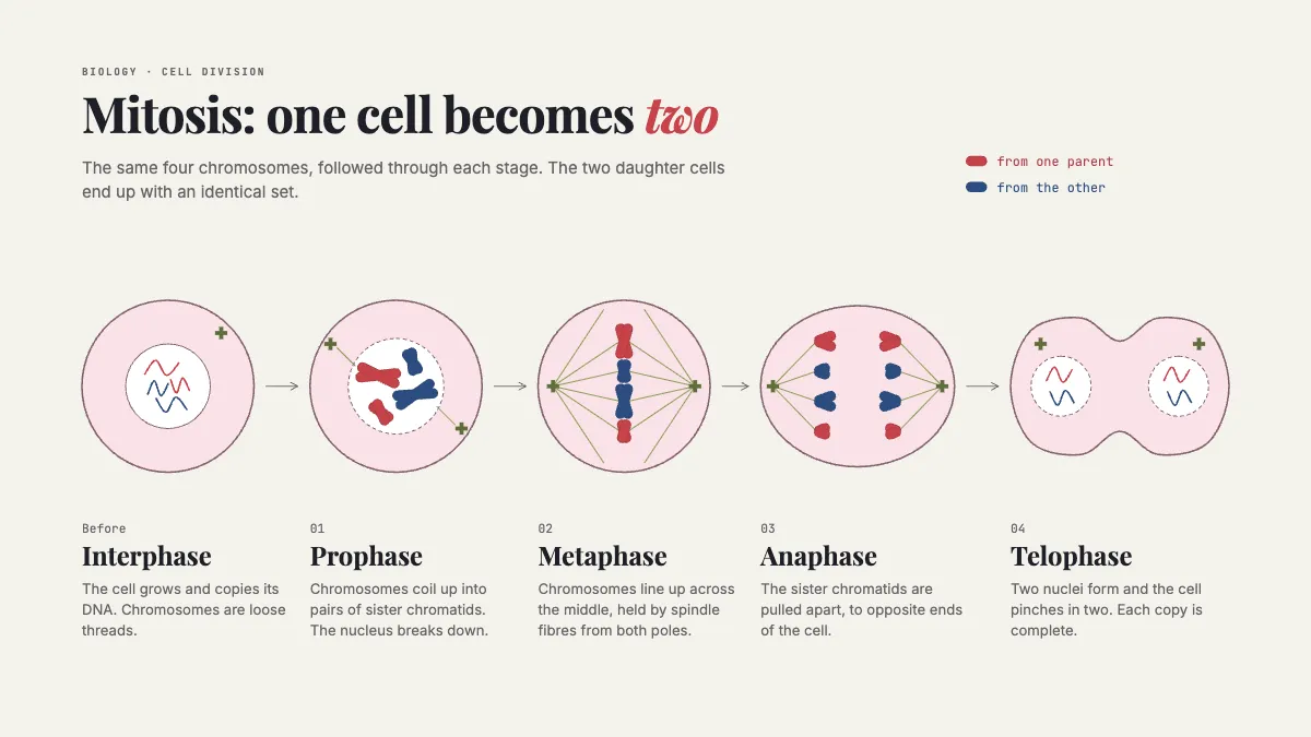
Interphase to telophase as five drawn cells, following the same four chromosomes from stage to stage.
Free. It opens in the Motionix editor as your own copy.
Download the pictureWebP, 1200 × 675, free to use. To change the words or colours, open it in the editor.
- Size
- 1920 × 1080 px
- Pages
- 1
- Motion
- Yes, plays live and in GIF or video
- Category
- Health and science
What it shows
- A row of drawn stages
- Chromatids that move apart
- One line per stage
Good for
How to use it
- Open the template. It becomes a project in your account.
- Change the words, colours and pictures. Every part is an ordinary shape, line or text box.
- Press Play to watch it build, then present it or export it.
What is a mitosis stages diagram?
A mitosis stages diagram shows one cell dividing into two identical cells as a sequence of drawn stages. The four phases of mitosis are prophase, metaphase, anaphase and telophase, followed by cytokinesis, when the cytoplasm splits. Interphase is usually drawn first, since that is when the cell grows and copies its DNA, although it is not itself part of mitosis.
When it helps
- An A level or GCSE biology lesson teaching the order of the phases.
- Revision before an exam question that asks students to identify a stage from a micrograph.
- Comparing mitosis with meiosis, using the same chromosomes in both diagrams.
- Explaining growth and repair, and why cancer is described as uncontrolled cell division.
Tips
- Use the mnemonic PMAT for the four phases, but teach interphase separately so students do not count it as a phase of mitosis.
- Keep each chromosome's colour constant across every stage; changing colours between drawings makes students think the chromosomes themselves change.
- Write one plain sentence per stage under its drawing, describing what moves, rather than a paragraph students will not read.
How to draw the stages of mitosis
Choose a small chromosome set
Use four chromosomes, two long and two short, and give them distinct colours. Following the same few chromosomes through every stage makes the copying visible.
Draw interphase
Show an intact nucleus with loose, thread-like chromatin. Add a note that the DNA is copied in this stage, ready for division.
Draw prophase and metaphase
In prophase, coil each chromosome into two sister chromatids joined at the centromere and break up the nuclear envelope. In metaphase, line them up across the middle, attached to spindle fibres.
Draw anaphase
Pull the sister chromatids apart toward opposite poles, with the centromeres leading and the arms trailing, so the direction of movement is clear.
Draw telophase and cytokinesis
Form a new nuclear envelope round each set and pinch the cell membrane in the middle. Each daughter cell gets the same four chromosomes.
FAQQuestions
Common questions.
What are the stages of mitosis in order?
Prophase, metaphase, anaphase and telophase, followed by cytokinesis, when the cell divides into two. Interphase comes before mitosis and is when the cell grows and copies its DNA. Some courses add prometaphase between prophase and metaphase.
What is the difference between mitosis and meiosis?
Mitosis makes two daughter cells genetically identical to the parent, with the full number of chromosomes, for growth and repair. Meiosis makes four genetically different cells with half the chromosome number, which become eggs or sperm.
What happens during cytokinesis?
Cytokinesis is the division of the cytoplasm that completes cell division. In animal cells the membrane pinches inward until the cell splits in two; in plant cells a new cell plate forms across the middle because the rigid wall cannot pinch.
More health and science templates
Patient guides, care pathways and the life sciences.