Free templateAnimal cell diagram
Inside an animal cell
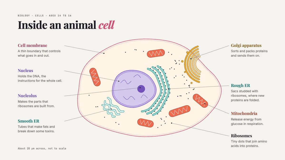
A fine-line animal cell with every organelle named in the margin, three colours for three systems.
Free. It opens in the Motionix editor as your own copy.
Download the pictureWebP, 1200 × 675, free to use. To change the words or colours, open it in the editor.
- Size
- 1920 × 1080 px
- Pages
- 1
- Motion
- Yes, plays live and in GIF or video
- Category
- Health and science
What it shows
- Drawn organelles
- Margin notes with leaders
- Colour by system
Good for
How to use it
- Open the template. It becomes a project in your account.
- Change the words, colours and pictures. Every part is an ordinary shape, line or text box.
- Press Play to watch it build, then present it or export it.
What is an animal cell diagram?
An animal cell diagram is a labelled drawing of a typical animal cell showing its main parts and what each one does. School diagrams usually name the cell membrane, cytoplasm, nucleus and mitochondria, and older students add the nucleolus, ribosomes, rough and smooth endoplasmic reticulum, the Golgi apparatus and vesicles. It is a simplified model: real cells vary widely in shape and size.
When it helps
- A GCSE biology lesson introducing organelles before students look down a microscope.
- Comparing animal and plant cells, side by side, in a revision session.
- A worksheet with the labels removed for students to complete as a test.
- Explaining how a protein is made and exported, following it from ribosome to membrane.
Tips
- Colour organelles by system, for example the nucleus and ribosomes for instructions and proteins, ER, Golgi and vesicles for transport, and mitochondria for energy, so function is visible at a glance.
- State the scale and that the drawing is not to scale; a typical animal cell is roughly 10 to 30 micrometres across.
- Never draw a cell wall, chloroplasts or a large permanent vacuole; examiners look for those mistakes.
How to draw and label an animal cell
Draw an irregular outline
Animal cells have no cell wall, so draw the membrane as a soft, uneven shape rather than the rigid rectangle used for plant cells.
Place the nucleus
Draw a large nucleus with a double-line envelope and a darker nucleolus inside. It does not need to sit exactly in the middle.
Add the membrane systems
Draw rough ER as folded sheets dotted with ribosomes next to the nucleus, smooth ER as tubes without dots, and the Golgi apparatus as a stack of curved sacs with vesicles.
Scatter mitochondria and ribosomes
Draw several oval mitochondria with folded inner membranes, and sprinkle free ribosomes through the cytoplasm as tiny dots.
Label with straight leader lines
Write labels in the margin and join each to its part with a ruled line that does not cross another. Add one line saying what each part does.
FAQQuestions
Common questions.
What are the parts of an animal cell?
The main parts are the cell membrane, cytoplasm, nucleus and mitochondria, plus ribosomes where proteins are made. More detailed diagrams add the nucleolus, rough and smooth endoplasmic reticulum, the Golgi apparatus, vesicles and lysosomes.
What is the difference between a plant cell and an animal cell?
Plant cells have three structures animal cells lack: a rigid cell wall made of cellulose, chloroplasts for photosynthesis, and a large permanent vacuole filled with cell sap. Both have a membrane, cytoplasm, nucleus, mitochondria and ribosomes.
What does the mitochondria do in an animal cell?
Mitochondria are where aerobic respiration takes place. They release energy from glucose using oxygen, and the cell uses that energy for everything from building proteins to muscle contraction. Cells that need a lot of energy, such as muscle cells, contain many of them.
More health and science templates
Patient guides, care pathways and the life sciences.